

DICOM annotation tool
for CT and MRI.
Open CT and MRI volumes from DICOM, NRRD and NIfTI files in axial, sagittal, coronal and 3D views. Segment anatomy as 3D masks, let AI and interpolation fill the slices in between, and review every volume in one place.
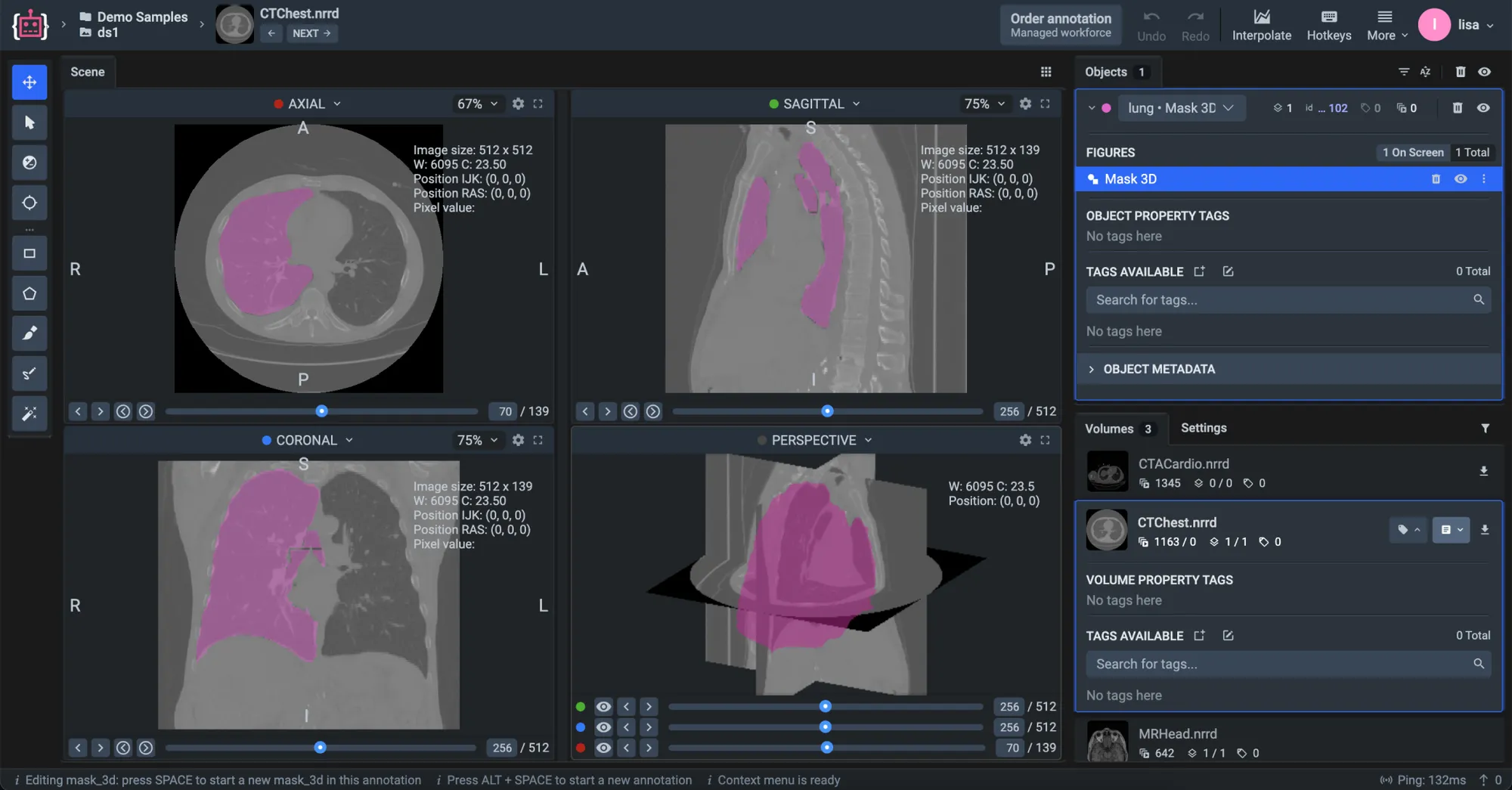
Every plane of a volume at once.
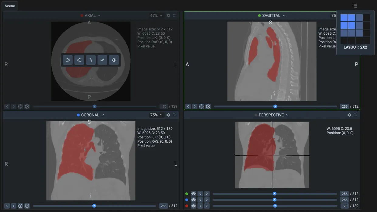
Every scan opens with its axial, sagittal, coronal and 3D views side by side, so a structure is labeled with its full anatomy in sight, not slice by slice in the dark.
Familiar to clinicians
The views follow the conventions of ITK-SNAP, 3D Slicer and MITK, with windowing presets, so doctors and other medical experts start with little to learn.
Every view in step
One click on the crosshair lines up every plane and the 3D view on the same point, and layouts grow up to 4 × 4 views.
Any source, one viewer
DICOM series, NRRD and NIfTI files open in the same views with the same tools, so mixed datasets need no conversion first.
Segment anatomy as true 3D masks that stay fast.
A lung or a tumor is one 3D object, not a stack of separate outlines. Draw it on any plane, and it updates on every other plane and in 3D.
One object, every plane
The brush, the pen and the Smart Tool all write into the same volumetric mask, whichever plane you work on.
Fast on full-size scans
3D masks load and decode quickly with far less memory, and the volume brush keeps up with every stroke.
Neighbours stay intact
Paint over everything, or only into voxels no other structure has claimed yet, so adjacent organs don't bleed into each other.
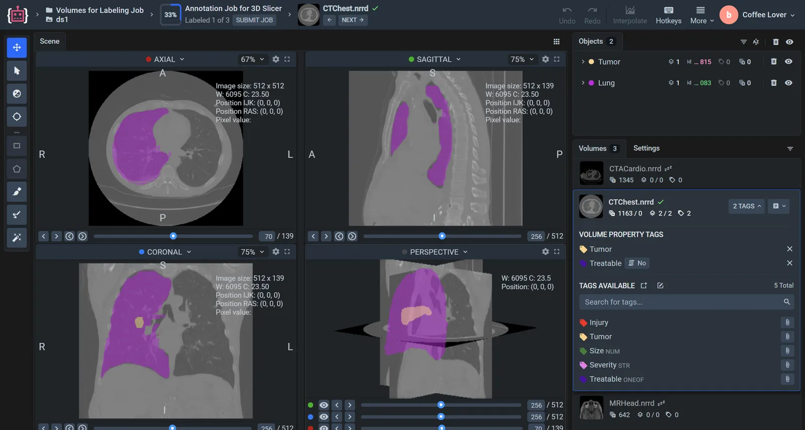
Label a few slices, and AI and interpolation do the rest.
A structure can span hundreds of slices. Annotators outline it on a handful of key slices, and the volume fills in between.
Segment a key slice with AI
Frame a structure with a box, and an interactive segmentation model draws its mask; a few clicks correct it. Serve a model trained on your own scans, and it works the same way.
Interpolate between slices
Interpolation matches the shapes on neighbouring labeled slices and reconstructs the object across the slices in between, in one plane or across all three.
Check it in 3D
The key slices and the result form one 3D mask. Review it in the 3D view and fix it on any plane.
Expert review without sending files around.
Annotators label, clinical experts review, and every volume stays in one project, with no exports passed back and forth.
Labeling tasks for volumes
Split a project between annotators with its classes and instructions, and send each volume to an expert who accepts it or returns it for another pass.
Throughput you can measure
Labeling Performance counts volumes too: time spent and acceptance rate per annotator.
Patient data protected
DICOM imports anonymize patient names and IDs, and the Enterprise edition runs on your own servers, even without internet access.
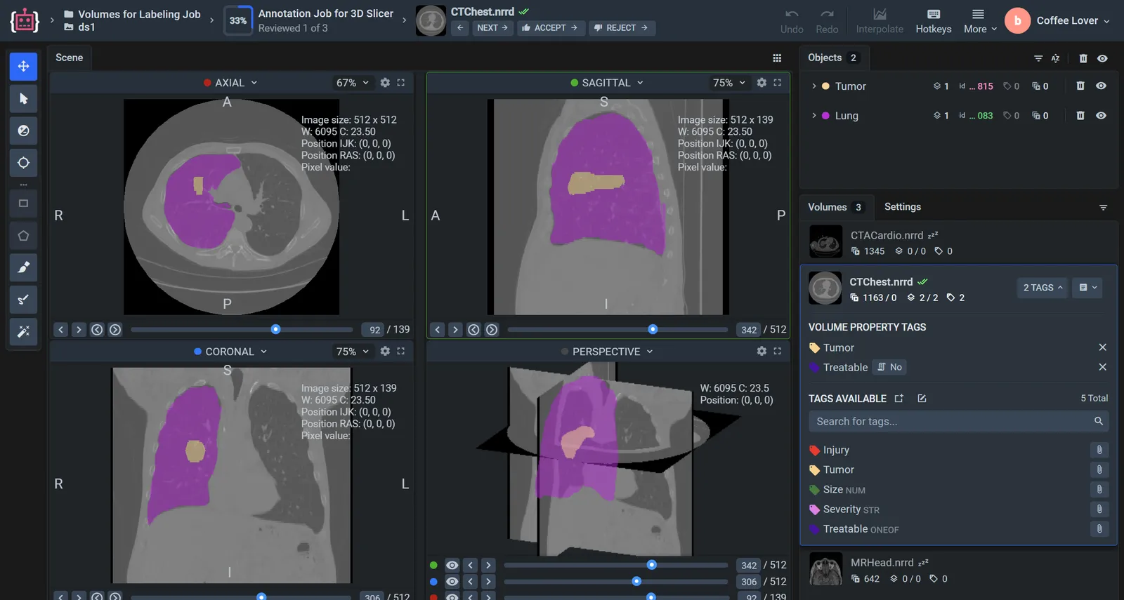
Annotators who live in 3D Slicer can stay there.
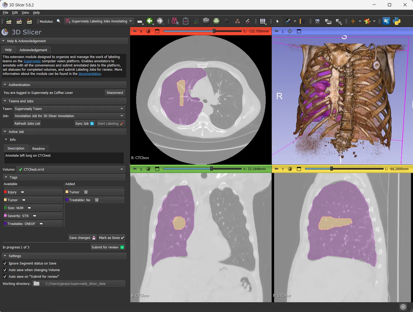
The ConnectToSupervisely extension opens Supervisely labeling tasks inside 3D Slicer. People label with 3D Slicer's own tools, and the results go back to the same project, ready for review in either app.
Connect
Install the extension from 3D Slicer's Extensions Catalog, sign in to Supervisely Cloud or your own instance, and pick a labeling task.
Label
Each class becomes a segmentation and each object a segment. Tag the volume and mark it as done.
Review
Submit the task for review. Reviewers accept or reject volumes in 3D Slicer or in Supervisely.
DICOM, NRRD and NIfTI in; open medical formats out.
Drop scans into a project and Supervisely builds the volumes. Labels leave in formats that 3D Slicer, MITK and training code read directly.
- .dcm
DICOM
Each series of slices becomes one volume, with patient names and IDs anonymized.
- .nrrd
NRRD
Single-file volumes, the format Supervisely keeps volumes in.
- .nii · .nii.gz
NIfTI
Volumes with their segmentation masks, imported as labels to correct.
- NRRD · NIfTI · STL
Export
3D masks as NRRD or NIfTI, per object or as one semantic mask, or as STL and OBJ surfaces.
Surgical videos go to the video labeling toolbox, with AI object tracking: see AI tools for surgical video annotation.
Frequently asked questions.
Which file formats does the DICOM annotation tool support?
Volumes in DICOM (.dcm series), NRRD (.nrrd) and NIfTI (.nii, .nii.gz), and projects in Supervisely format. NIfTI files can bring segmentation masks along as labels. Single DICOM, NRRD and NIfTI images open in the image labeling toolbox. See the volume formats in the docs.
Can I label CT and MRI scans in 3D?
Yes. Every volume opens in axial, sagittal, coronal and 3D views, and 3D masks are drawn and edited on any plane. Label a few key slices, and interpolation reconstructs the object across the slices in between. Read the guide to DICOM and NIfTI annotation.
Which AI models help with medical image segmentation?
The Smart Tool runs interactive segmentation models, such as Segment Anything, on the slice you label: frame a structure with a box and correct the mask with clicks. You can switch between models or serve one you trained on your own scans. See AI-assisted labeling.
How is patient data protected?
DICOM imports anonymize patient names and IDs by default. Team roles control who sees each project, and the Enterprise edition runs on your own servers, even without internet access. See security and permissions.
Does Supervisely work with 3D Slicer?
Yes. With the ConnectToSupervisely extension, annotators and reviewers work on Supervisely labeling tasks inside 3D Slicer, and exported NRRD volumes and masks open in 3D Slicer and MITK as they are. See how to use 3D Slicer with Supervisely.